Monday, April 6, 2020

Optochin Susceptibility Test for Streptococcus pneumoniae

Optochin Susceptibility Test for Streptococcus pneumoniae

Streptococcus pneumoniae is found commonly in human respiratory tract, as other streptococci, and has a hemolytic pattern indistinguishable from that of other alpha-hemolytic streptococci and Lactococci.
Optochin (ethylhydrocupreine hydrochloride) is a chemical (quinine derivative) that is used in the presumptive identification of alpha-hemolytic Streptococcus pneumoniae, which is optochin sensitive. Other alpha-hemolytic Streptococcal species are resistant to optochin. (Mnemonic: OVRPS: Optochin- Viridians Resistant and Optochin- Pneumococcus sensitive). Optochin is completely soluble in water.
The optochin test is widely used in the form of filter paper discs. In 1955 Bowen and Jeffries impregnated disks with the reagent ethylhydrocupreine hydrochloride, which are applied directly to inoculated plates before incubation to demonstrate the susceptibility of pneumococcus for identification purpose.

Principle of Optochin Susceptibility Test

Optochin Susceptibility test is used to determine the effect of optochin reagent (<5 a="" agar="" an="" blood="" diffusion="" disk="" em="" g="" is="" medium="" ml="" nbsp="" on="" optochin="" organism.="" performed="" principle.="" sensitive="" style="box-sizing: border-box;" test="" this="" using="">S. pneumoniae 
surrounding the disc are lysed due to the chemical optochin that changes in surface tension, resulting in zone of inhibition.

Reagents

Optochin disks- each disk is impregnated with 5µg of optochin.5% sheep blood agar

Procedure

  1. Using an inoculating loop, select a well-isolated colony of the alpha-hemolytic organism to be tested.
  2. Streak the isolates onto 5%sheep blood agar plate.
    (NOTE: Use of media other than 5% sheep blood agar is not recommended, as smaller zone sizes can result in lack of definitive identification)
  3. Using sterile forceps, place an optochin disk onto the inoculated surface of the agar.
  4. Press disk gently with the sterile forceps or loop so that the disk adheres firmly to the agar surfaces.
  5. Incubate the plate at 35-37°C fir 18-24 hour in 5 to 10% CO2.
    (NOTE: Cultures do not grow as well in ambient air, and larger zone of inhibition occur.)
  6. Measure the diameter of zone of inhibition including diameter of disk.

Result interpretation
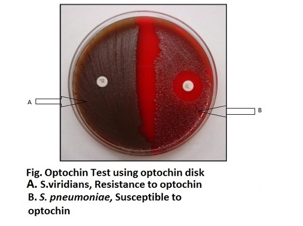
Result Interpretation of Optochin test
Sensitive: zone of inhibition ≥ 14mm (15-30mm) around the 6mm disk.
(NOTE: For 10mm disk, use ≥ 16mm zone of inhibition.
If the inhibition zone is less than 14 mm, further testing (bile solubility or serology) is indicated for the identification of S. pneumoniae.)
Intermediate: Organisms with zone of inhibiton < 14mm around the disk. The strain is identified as pneumococcus only if it is bile soluble.
Resistant: No zone of inhibition around the disk.

Sunday, April 5, 2020

Antibiotic resistance

What is antimicrobial resistance?
Antimicrobial resistance (AMR), or drug resistance, develops when microbes, including bacteria, fungi, parasites, and viruses, no longer respond to a drug that previously treated them effectively.
AMR can lead to the following issues:
  • some infections being harder to control and staying longer inside the body
  • longer hospital stays, increasing the economic and social costs of infection
  • a higher risk of disease spreading
  • a greater chance of fatality due to infection
A significant concern is that AMR could lead to a post-antibiotic era in which antibiotics would no longer work.
This would mean that common infections and minor injuries that became straightforward to treat in the 20th century could again become deadly.

Antibiotic versus antimicrobial resistance

Distinguishing between antibiotic and antimicrobial resistance is important.
  • Antibiotic resistance refers to bacteria resisting antibiotics.
  • Antimicrobial resistance (AMR) describes the opposition of any microbe to the drugs that scientists created to kill them.
It is possible for AMR to develop in bacteria, but it can also originate in fungi, parasites, and viruses. This resistance could affect people with Candida, malaria, HIV, and a wide range of other conditions.

Microbes can become resistant to drugs for both biological and social reasons.

Microbial behavior

As soon as scientists introduce a new antimicrobial drug, there is a good chance that it will become ineffective at some point in time.
This is due primarily to changes occurring within the microbes.
These changes can come about in different ways:
Mutation: When microbes reproduce, genetic mutations can occur. Sometimes, this will create a microbe with genes that help it survive in the face of antimicrobial agents.
Selective pressure: Microbes that carry these resistance genes survive and replicate. The newly generated resistant microbes eventually become the dominant type.
Gene transfer: Microbes can pick up genes from other microbes. Genes conferring drug resistance can easily transfer between microbes.
Phenotypic change: Microbes can change some of their characteristics to become resistant to common antimicrobial agents.

People’s behavior

The way in which people use antimicrobial drugs is a significant contributing factor. For example:
Inexact diagnosis: Doctors sometimes prescribe antimicrobials “just in case,” or they prescribe broad-spectrum antimicrobials when a specific drug would be more suitable. Using these medications in this way increases the risk of AMR.
Inappropriate use: If a person does not complete a course of antimicrobial drugs, some microbes may survive and develop resistance to the drug.
Resistance can also develop if people use drugs for conditions that they cannot treat. For example, people sometimes take an antibiotic for a viral infection.
Agricultural use: Using antibiotics in farm animals can promote drug resistance. Scientists have found drug-resistant bacteria in meat and food crops that have exposure to fertilizers or contaminated water. In this way, diseases that affect animals can pass to humans.
Hospital use: People who are critically ill often receive high doses of antimicrobials. This encourages the spread of AMR microbes, particularly in an environment where various diseases are present.
The United States Food and Drug Administration (FDA) point out that doctors often give antibiotics as a treatment for a sore throat. However, only 15 percent of sore throats are due to streptococcal bacteria. In many cases, antibiotics cannot cure a sore throat.
The FDA add that doctors write “tens of millions” of prescriptions for antibiotics that offer no benefit each year.
People who use these drugs are at risk of allowing AMR to develop. This could make them more likely to have a health problem in the future that will not respond to antibiotics.

Antimicrobial resistance can occur in bacteria, viruses, fungi, and parasites.
Below are some examples:
Tuberculosis (TB): This airborne lung disease results from a bacterial infection. TB was a major killer before antibiotics became available. More recently, drug-resistant forms of TB have emerged worldwide. Standard antibiotic treatments will not work against these forms of the disease.
A person who has TB that is not drug-resistant will require daily treatment with several drugs for 6 to 9 months.
Drug-resistant TB is more complex to treat. The person will need to take the drugs for a longer time, and they will need close supervision. Poor management can result in fatalities.
Methicillin-resistant Staphylococcus aureus (MRSA): This is a bacterial infection that can be fatal. People usually get MRSA when they are staying in a hospital.
In the past, it was a well-controlled infection, but now the CDC see it as a major public health concern due to antibiotic resistance.
Gonorrhea: Gonorrhea is a sexually transmitted bacterial infection that is common in the U.S. and elsewhere. Cases of drug-resistant gonorrhea have started to occur.
Now, there is only one type of drug that is still effective against the drug-resistant form of this disease.
The CDC describe drug-resistant gonorrhea as an “urgent public health threat.”
Escherichia coli (E. coli): This bacterium is a common cause of food-borne disease and urinary tract infections. The rate of antibiotic resistance in E. coli is increasing quickly.
HIV: Effective antiviral treatment for HIV can now prevent this condition from becoming more severe. The treatment can make the levels of the virus undetectable, meaning that it is not transmissible.
The World Health Organization (WHO) note that if people are unable to take the drugs as they should, perhaps due to medical costs, new drug-resistant strains of the virus may appear.
Fungal infections: Candida, Aspergillus, and other fungi can lead to a range of severe infections. Candida albicans (C. albicans) is responsible for thrush, a common vaginal infection. Aspergillus can cause or worsen aspergillosis, a lung condition.
Some of these infections can have fatal consequences. There is concern that fungi are becoming increasingly resistant to antimicrobial treatments.
Malaria: Mosquitoes spread this parasitic disease, which killed around 445,000 people worldwide in 2016. In many parts of the world, drug-resistant parasites have evolved so that certain antimalarial drugs are now ineffective.

As infections stop responding to current drugs, there is an urgent need to find alternatives.
In some cases, this means using combinations of different medications, known as multiple-drug therapy.
Scientists are also looking for new forms of treatment, including different types of antibiotics and other alternatives.

What are the alternatives?

Scientists have proposed some novel ways of combating bacteria.
These include the following techniques, which researchers are investigating for the treatment of Clostridium difficile (C. difficile):
  • using a virus that consumes bacteria, known as a bacteriophage, in drug form
  • using monoclonal antibodies that can combat the effects of the toxins that the microbes produce
  • developing vaccines to prevent infection from occurring
  • fecal microbiota transplant, which involves taking good bacteria from a healthy person’s gut and transplanting them into a recipient who is lacking them
  • the use of probiotics to restore the gut flora
More research into these treatments is necessary to confirm their effectiveness.
Meanwhile, experts are stressing the need for:
  • doctors to prescribe antibiotics only when they are useful and necessary
  • patients to use antimicrobial drugs precisely as the doctor recommends and only after a complete diagnosis

hand washingShare on Pinterest
Preventing diseases from spreading, for example, through good hygiene, is one way to reduce the need for or use of medications.
Preventing microbes from developing resistance to drugs has become as important as treating the illnesses that they cause.
The main reason for the increase in AMR appears to be the frequent and improper use of antimicrobial drugs.
Steps that people can take to help lower the risk of AMR include the following:
  • Only use antimicrobial drugs when a doctor prescribes them.
  • Always complete the full prescribed course, even if the symptoms have subsided. If not, the drug may only kill off the most vulnerable microbes, leaving others to survive and develop resistance.
  • Never share antimicrobials with others or using leftover drugs from previous prescriptions. These medications may not be suitable for different forms of infection.
  • Do not pressurize doctors into prescribing antimicrobials when they are not necessary.
  • Follow good hygiene practices to prevent the spread of microbes, including washing hands thoroughly and ensuring that food preparation areas are clean.
  • Get recommended vaccinations, as this will reduce the risk of needing to take medication.

Antibiotic Susceptibility Testing

Objective


The test determines the susceptibility of a microbial species against different antibiotic agents.

Principle


The introduction of various antimicrobials for treating variety of infections showed the necessity of performing antimicrobial susceptibility testing as a routine procedure in all microbiology laboratories. In laboratories it can be made available by using antibiotic disk which will diffuse slowly into the medium where the suspected organism is grown. The basic principle of the antibiotic susceptibility testing has been used in microbiology laboratories over 80 years. Various chemical agents such as antiseptics, disinfectants, and antibiotics are employed to combat with the microbial growth. Among these, antibiotics are generally defined as the substances produced by the microorganism such as Penicillium, which has the ability to kill or inhibit the growth of other microorganisms, mainly bacteria. Antimicrobial susceptibility tests (ASTs) basically measures the ability of an antibiotic or other antimicrobial agent to inhibit the invitro microbial growth.

There are many different procedures that microbiologists use to study the effects of various antimicrobial agents in treating an infection caused by different microorganisms.
Mueller Hinton Agar is considered as best for the routine susceptibility testing since it is has batch-to-batch reproducibility, low concentration of inhibitors of sulphonamide, trimethoprim and tetracyclines and produce satisfactory results for most of the non-fastidious pathogens. Fastidious organisms which require specific growth supplements need different media to grow for studying the susceptibility patterns.

The Kirby Bauer test is a qualitative assay whereby disks of filter paper are impregnated with a single concentration of different antibiotics or any chemicals that will diffuse from the disk into the agar. The selected antibiotic disks are placed on the surface of an agar plate which has already been inoculated with test bacteria. During the incubation period, the antibiotics/chemicals diffuse outward from the disks into the agar. This will create a concentration gradient in the agar which depends on the solubility of the chemical and its molecular size. The absence of growth of the organism around the antibiotic disks indicates that, the respected organism is susceptible to that antibiotic and the presence of growth around the antibiotic disk indicates the organism is resistant to that particular antibiotic. This area of no growth around the disk is known as a zone of inhibition, which is uniformly circular with a confluent lawn of growth in the media.
The diameters of the zone of inhibition are measured (including disk) using a metric scale or a sliding caliper. The measured zone diameter can be compared with a standard chart for obtaining the susceptible and resistant values. There are zone of intermediate resistance which means that the antibiotic may not be sufficient enough to eradicate the organism from the body.


Examples of Antibiotic Sensitivity Testing Methods

1. DILUTION METHODS


The Broth dilution method involves subjecting the isolate to a series of concentrations of antimicrobial agents in a broth environment.   Microdilution testing uses about 0.05 to 0.1 ml total broth volume and can be conveniently performed in a microtiter format.  Macrodilution testing uses broth volumes at about 1.0 ml in standard test tubes.  For both of these broth dilution methods, the lowest concentration at which the isolate is completely inhibited (as evidenced by the absence of visible bacterial growth) is recorded as the minimal inhibitory concentration or MIC.  The MIC is thus the minumum concentration of the antibiotic that will inhibit this particular isolate.  The test is only valid if the positive control shows growth and the negative control shows no growth.

A procedure similar to broth dilution is agar dilution.  Agar dilution method follows the principle of establishing the lowest concentration of the serially diluted antibiotic concentration at which bacterial growth is still inhibited. 

2-23-DISK-DIFFUSION.jpgOn this agar plate, a bacterial isolate is tested for resistance to each of twelve different antibiotics.  The clear zones around each disc are the zones of inhibition that indicate the extent of the test organism’s inability to survive in the presence of the test antibiotic. (A)The disk shows a large zone of inhibition; whereas (B) shows no zone of inhibition, indicating resistance of the isolate to the test antibiotic 

Presence of zone of inhibition is not automatically interpreted as susceptibility to the antibiotic; the zone width has to be measured and compared against a reference standard which contains measurement ranges and their equivalent qualitative categories of susceptible, intermediately susceptible or resistant.

2-24-DISK-DIFFUSION-CLOSE-U.jpgFor example, this E.coli isolate on the right has a zone of inhibition of 10.1mm around ampicillin (AM); since the zone diameter interpretation chart is as follows:
Resistant: 13mm or less
Intermediate: 14-16 mm
Susceptible: 17 mm or more
This particular E.coli isolate is read as resistant to ampicillin.

2. DISK DIFFUSION METHOD


Because of convenience, efficiency and cost, the disk diffusion method is probably the most widely used method for determining antimicrobial resistance in private veterinary clinics. 

A growth medium, usually Mueller-Hinton agar, is first evenly seeded throughout the plate with the isolate of interest that has been diluted at a standard concentration (approximately 1 to 2 x 10⁸ colony forming units per ml).  Commercially prepared disks, each of which are pre-impregnated with a standard concentration of a particular antibiotic, are then evenly dispensed and lightly pressed onto the agar surface.  The test antibiotic immediately begins to diffuse outward from the disks, creating a gradient of antibiotic concentration in the agar such that the highest concentration is found close to the disk with decreasing concentrations further away from the disk. After an overnight incubation, the bacterial growth around each disc is observed.  If the test isolate is susceptible to a particular antibiotic, a clear area of “no growth” will be observed around that particular disk.

The zone around an antibiotic disk that has no growth is referred to as the zone of inhibition since this approximates the minimum antibiotic concentration sufficient to prevent growth of the test isolate.  This zone is then measured in mm and compared to a standard interpretation chart used to categorize the isolate as susceptible, intermediately susceptible or resistant.  MIC measurement cannot be determined from this qualitative test, which simply classifies the isolate as susceptible, intermediate or resistant.


3. E-TEST


E-test (AB Biodisk, Solna, Sweden) is a commercially available test that utilizes a plastic test strip impregnated with a gradually decreasing concentration of a particular antibiotic.  The strip also displays a numerical scale that corresponds to the antibiotic concentration contained therein. This method provides for a convenient quantitative test of antibiotic resistance of a clinical isolate.  However, a separate strip is needed for each antibiotic, and therefore the cost of this method can be high.

4. AUTOMATED ANTIMICROBIAL SUSCEPTIBILITY TESTING SYSTEMS


Several commercial systems have been developed that provide conveniently prepared and formatted microdilution panels as well as instrumentation and automated reading of plates.  These methods are intended to reduce technical errors and lengthy preparation times.

Most automated antimicrobial susceptibility testing systems provide automated inoculation, reading and interpretation.  These systems have the advantage of being rapid (some results can be generated within hours) and convenient, but one major limitation for most laboratories is the cost entailed in initial purchase, operation and maintenance of the machinery.  Some examples of these include: Vitek System (bioMerieux, France), Walk-Away System (Dade International, Sacramento, Calif.), Sensititre ARIS (Trek Diagnostic Systems, East Grinstead, UK), Avantage Test System (Abbott Laboratories,  Irving, Texas), Micronaut (Merlin, Bornheim-Hesel, Germany), Phoenix  (BD Biosciences, Maryland) and many more. 


5. MECHANISM-SPECIFIC TESTS


Resistance may also be established through tests that directly detect the presence of a particular resistance mechanism.  For example, beta lactamase detection can be accomplished using an assay such as the chromogenic cephalosporinase test (Cefinase disk by BD Microbiology Systems, Cockeysville, MD and BBL DrySlide Nitrocefin, Becton Dickinson, Sparks, MD) and detection for chloramphenicol modifying enzyme chloramphenicol acetyltransferase (CAT) may utilize commercial colorimetric assays such as a CAT reagent kit (Remel, Lenexa, Kansas).


Materials Required


  1. Petriplate containing microbial culture(For example, Escherichia coli)
  2.  Inoculation loop
  3. Bunsen burner
  4. Saline solution
  5. McFarland solution
  6. MHA plate
  7. Cotton swab
  8. Antibiotic disks(Streptomycin (S), Ciprofloxacin (CIP), Chloramphenicol (C), Doxycycline (D), Penicillin G (P), Gentamycin (G)
  9. Tooth pick
  10. Incubator
  11. Ruler
     

Procedure


  1. Select a pure culture plate of one of the organisms to be tested.
  2. Aseptically emulsify a colony from the plate in the sterile saline solution. Mix it thoroughly to ensure that no solid material from the colony is visible in the saline solution.
  3. Repeat until the turbidity of the saline solution visually match that of the standard turbidity.
  4. Take a sterile swab and dip it into the broth culture of organism.
  5. Gently squeeze the swab against the inside of the tube in order to remove excess fluid in the swab.
  6. Take a sterile Mueller-Hinton agar (MHA) plate or a nutrient agar (NA) plate.
  7. Use the swab with the test organism to streak a MHA plate or a NA plate for a lawn of growth.
  8. After the streaking is complete, allow the plate to dry for 5 minutes.
  9. Antibiotic discs can be placed on the surface of the agar using sterilized forceps.
  10. Gently press the discs onto the surface of the agar using flame sterilized forceps or inoculation loop.
  11. Carefully invert the inoculated plates and incubate for 24 hours at 37° C.
  12. After incubation, use a metric ruler to measure the diameter of the zone of inhibition for each antibiotic used.
  13. Compare the measurement obtained from the individual antibiotics with the standard table to determine the sensitivity zone.
  14. Compare the measurement obtained from the individual antibiotics to the standard table to determine whether the tested bacterial species is sensitive or resistant to the tested antibiotic.


Antimicrobial therapy


Antimicrobial therapy


Terminology
§  Chemotherapy - use of chemical agents to treat disease
§  Chemotherapeutic agents - chemical agents used for treatment
§  Antimicrobial agent - chemical agent used to treat diseases caused by microbes
Three groups of antimicrobial agents:
v  Synthetic agents - produced in laboratory
v  Natural agents - metabolic products (secondary metabolites) produced by certain groups of fungi and fungal-like bacteria that are antibacterial in action; commonly called antibiotics.
v  Semi-synthetic agents - derivatives of natural agents altered in laboratory by adding chemical groups (to improve effectiveness, etc.)
Criteria that determines the effectiveness of antimicrobial agents used in the treatment of infectious diseases:
ü  Selective toxicity - destroys or inhibits microbe without affecting host cells
ü  Broad spectrum - effective against a wide variety of organisms
ü  Non-mutagenic - does not induce development of resistant strains
ü  Soluble in body fluids - distributed through body (in bloodstream)
ü  Stable in body fluids - not easily broken down or excreted, to maintain constant and effective levels
ü  Absorbed by tissues - to reach site of infection
ü  Non-allergenic to host - should not cause adverse reactions in host
ü  Should not disturb host’s normal flora (organisms normally living in body) causing secondary (super) infections produced by opportunists
Modes of action
ž  Interfere with microbe’s vital metabolic process that does not occur in host cells (inhibit microbial enzymes) Act by: a.Competitive inhibition – competes with essential substrate to act with microbial enzyme.  b. Non-competitive inhibition – reacts directly with enzyme

ž  Targets structural/ metabolic differences between eukaryotic, prokaryotic cells.

ž  Action of antimicrobial agents - interfere with:
o   Metabolic pathways – production of an essential metabolite (By competitive inhibition)
o   The cell wall (murein) synthesis.
o   Protein synthesis (enzymes)
o   Nucleic acid replication/transcription.
o   Cause damage to cell membrane
Examples of antimicrobial agents and their actions 
Ø  Sulfonamides - Metabolic Pathway - Inhibits synthesis folic acid by competitive inhibition.
Ø   Penicillin - inhibits Cell Wall Synthesis
Ø  Erythromycin – Stops Protein Synthesis
Ø  Tetracyclines  -  Stops protein synthesis
Ø   Chloramphenicol  -  Stops protein synthesis
Ø  Streptomycin -  Stops protein synthesis
Ø  Quinolones  -  Inhibits DNA  Synthesis 
Ø  Rifampin  - Inhibits RNA Synthesis
Ø  AZT  -  Inhibits reverse transcriptase ad DNA Synthesis
Ø  Acyclovir, Ribavirin  - Prevents viral replication and  DNA, RNA Synthesis
Ø  Griseofulvin  -  affects RNA Synthesis
Ø  Polymixins  -  Damage Cytoplasmic Membrane
Disadvantages of Antimicrobial Therapy:
·         Allergic reactions
·         Toxic effects on normal tissues
·         Disturbs host normal flora ® secondary infections
·         Development of resistant strains – spontaneous mutations, DNA transfer